First Medial Cuneiform, Http Www Intjmorphol Com Wp Content Uploads 2018 12 Art 37 364 Pdf
First medial cuneiform Indeed recently has been hunted by users around us, perhaps one of you personally. Individuals now are accustomed to using the internet in gadgets to view video and image information for inspiration, and according to the name of the article I will discuss about First Medial Cuneiform.
- 6 Normal Variants And Anomalies Musculoskeletal Key
- Tarsometatarsal Arthritis Podiatry Orthopedics Physical Therapy
- 1
- Cuneiforms Cuneiform Bones Tarsal Bones Tarsus Foot Bones Bones Of The Feet Foot Regions Pedal Region Wellness Advocate Com
- Medial Cuneiform Bone High Resolution Stock Photography And Images Alamy
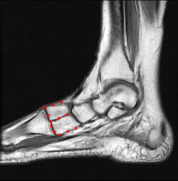
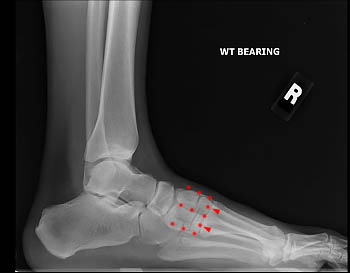
- Bipartite Medial Cuneiform Radsource
Find, Read, And Discover First Medial Cuneiform, Such Us:
- Dislocation Intermediate Cuneiform With Fracture Medial Cuneiform Journal Of Orthopaedic Case Reports
- What Is The Medial Cuneiform With Pictures
- Amicus Illustration Of Amicus Surgery Foot Cannulated Screw First Metatarsal Medial Cuneiform Navicular Midfoot Third Bone
- Bipartite Medial Cuneiform Radsource
- Anatomyexpert Medial Cuneiform Structure Detail
If you are looking for Remove Bg you've come to the ideal place. We ve got 104 images about remove bg including images, photos, photographs, wallpapers, and more. In these web page, we also provide variety of graphics available. Such as png, jpg, animated gifs, pic art, symbol, black and white, transparent, etc.

What Is The Medial Cuneiform With Pictures Remove Bg
Bipartite Medial Cuneiform Radsource Remove Bg
Http Www Intjmorphol Com Wp Content Uploads 2018 12 Art 37 364 Pdf Remove Bg
Http Www Podiatryinstitute Com Pdfs Update 1995 1995 36 Pdf Remove Bg
Https Encrypted Tbn0 Gstatic Com Images Q Tbn 3aand9gcskw3hyfvk5 E2olxwmml 3ga4rsiczqz8s Egniqb2z0bf9qqy Usqp Cau Remove Bg
Https Encrypted Tbn0 Gstatic Com Images Q Tbn 3aand9gcr5zgizwigqhed7 Akiu Dq2gqspjo7gjmhmoq9koract7i6vqw Usqp Cau Remove Bg
Its proximal articular surface is large and kidney shaped.
Remove bg. The medial cuneiform also known as first cuneiform is the largest of the cuneiforms. It is situated at the medial side of the foot anterior to the navicular bone and posterior to the base of the first metatarsal. The medial approach to the first metatarsal is used for the fixation of certain fractures of the first metatarsal for osteotomies of the first metatarsal or for transfer of the flexor hallucis longus tendon into the base of the first metatarsal.
In the adult the first metatarsal is supplied by an artery entering the first metatarsal head via the plantar distal metaphysis. Lateral to it is the intermediate cuneiform. The medial cuneiform is one of the cuneiforms it is the most medial in the distal row of tarsal bones.
It has a narrow dorsal surface and a flat plantar surface which receives a slip from the tibialis posterior tendon. Also known as the first cuneiform the medial cuneiform is the biggest cuneiform bone. The distal surface is reniform congruent with the articulating base of first metatarsal and proximal surface has a pyriform facet for the navicular.
The lateral cuneiform also articulates laterally with the cuboid in a variable manner. It is on the inner side of the foot behind the first metatarsal which leads to the bones of. Two muscles tibialis anterior and peroneus longus insert at the medial cuneiform bone.
Anatomically the medial cuneiform is positioned between the navicular bone in the ankle and the metatarsals in the front of the foot. Its circumference is grooved for the tarsometatarsal ligaments and medially gives insertion to part of the tendon of the tibialis anterior. It articulates with the first and second metatarsals the long bones connecting to the toes along with the navicular bone and the second cuneiform.
All three medial middle and lateral articulate with the navicular proximally. The cuneiform from the latin for wedge bones are a set of three bones in the medial side of the foot that articulate with the navicular proximally and with the proximal surfaces of metatarsal 1 3 distally. The medial cuneiform is found on the inside of the foot and connects to the first metatarsal.
Lateral third mt. The first metatarsal articulates forms joints with the medial cuneiform and to a small extent with the intermediate cuneiform bone. The navicular intermediate cuneiform as well as the first and second metatarsal bones.
Each articulates with a single metatarsal mt distally medial. The cuneiforms are located on the medial side of the midfoot. The medial cuneiform also called the first or internal cuneiform is located around the middle foot.
A change in the first ima after first metatarsophalangeal joint fusion showed a strong positive correlation with a change in cuneiform obliquity p 0001.
Bipartite Medial Cuneiform Radsource Remove Bg

Aneurysmal Bone Cyst Of Medial Cuneiform And A Novel Surgical Technique For Mid Foot Reconstruction Bmj Case Reports Remove Bg

Metatarsophalangeal Joint Pain Musculoskeletal And Connective Tissue Disorders Merck Manuals Professional Edition Remove Bg
Https Journals Sagepub Com Doi Pdf 10 1177 1071100718789696 Remove Bg
More From Remove Bg
- Fantastic Beasts All Movies List
- Fantastic Beasts And Where To Find Them Kowalski
- Fantastic Beasts And Where To Find Them 2 Zoe Kravitz
- Karib Tenger Kalozai Hbo Go
- Fantastic Beasts And Where To Find Them 3 Cast
Incoming Search Terms:
- Cuneiforms Cuneiform Bones Tarsal Bones Tarsus Foot Bones Bones Of The Feet Foot Regions Pedal Region Wellness Advocate Com Fantastic Beasts And Where To Find Them 3 Cast,
- File Medial Cuneiform Bone05 Inferior View Png Wikimedia Commons Fantastic Beasts And Where To Find Them 3 Cast,
- First Metatarsal And Medial Cuneiform Fantastic Beasts And Where To Find Them 3 Cast,
- Tarsals Metatarsals Phalanges Quiz Flashcards Quizlet Fantastic Beasts And Where To Find Them 3 Cast,
- Emdocs Net Emergency Medicine Educationfoot Injuries In The Emergency Department Emdocs Net Emergency Medicine Education Fantastic Beasts And Where To Find Them 3 Cast,
- Parts Of The Foot Diagram Quizlet Fantastic Beasts And Where To Find Them 3 Cast,